Humans share 96% of genetic heritage with their closest phylogenetic cousin, the chimpanzee. The existence of non-invasive brain imaging methods such as MRI has made it possible to carry out studies aimed at exploring the brain structures of chimpanzees in order to better understand their uniqueness. To date, diffusion MRI, which reports the microstructure of brain tissues by following the anisotropy of the Brownian motion of water molecules, remains the only method for in vivo imaging of the brain structural connectivity. Brain anatomy and connectivity are closely linked and major brain functions result from the establishment of functional networks made up of groupings (or bundles) of axonal fibers of varying length, populating the cerebral white matter. In a recent study published in NeuroImage, Baobab's team established the first complete atlas of chimpanzee brain connectivity from diffusion MRI images for which innovative computer processing had enabled the creation of two atlases of brain connectivity white matter, deep and superficial (see Joliot news).
If the mapping of long-distance connections is easier to establish, because their trajectories are little impacted by the folds and convolutions of the brain, the short white matter bundles (SWMBs) are impacted over their entire length, and therefore, constitute a relatively underexplored aspect of brain connectivity. The delimitation between deep beams and short beams remains a non-consensual debate within the scientific community.
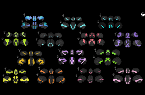
In this work, the team undertook a complete examination of the connectivity of SWMBs in human and chimpanzee brains, using a novel combination of empirical and geometric methodologies to objectively classify their morphology. It thus aims to discover the commonalities and disparities in the superficial white matter bundles between chimpanzees and humans. For this, authors relied on two anatomical atlases: the Ginkgo Chauvel chimpanzee atlas, established from a cohort of 39 chimpanzees and the Ginkgo Chauvel human atlas, specially designed for this study, established in 39 subjects, intentionally identical in gender and number with the cohort of chimpanzees. Each atlas has 844 and 1375 surface bundles, respectively, and the researchers focused on sparse representations of SWMB morphology. U-shaped fibers, already well described in humans, have also been observed in chimpanzees and other shapes, with more complex geometry, such as 6 and J shapes, were observed for the first time. A clustering approach based on an algorithm called isomap, originally developed for the study of sulco-gyral morphometry, allowed the authors to observe and locate several bundle morphologies, different between humans and chimpanzees.

Comparison of chimpanzee and human atlases by superficial white matter beams © M.Chauvel / CEA
One of the challenges of evolutionary neurosciences is to create and compare models of brain connectivity between different species. This study, which observes both commonalities and disparities in the SWMBs of chimpanzees and humans, with emphasis on potential relationships with cerebral gyrification, sheds light on the evolution and organization of these crucial neurons and should ultimately contribute to a better understanding of the evolution and cognitive development of humans.
Contact : Cyril Poupon (cyril.poupon@cea.fr)
- The white matter is made up of millions of myelinated axons, connecting neurons from one region of the brain to another, whose role is to transport nerve impulses. The organization of white matter is a guarantor of the proper functioning of the brain.
- Isomap is a dimension reduction algorithm that preserves the overall structure