You are here : Home > Research Centers and Units > MIRCen > MIRCen facilities > Clinical imaging

Clinical imaging

​​Les plateformes de MIRCen
​Platform's contact IC-MIRCen@cea.fr

Published on 3 July 2024

MIRCen's clinical activities are carried out in close collaboration with :

  1. ​The ​SHFJ's clinical imaging center, 
  2. ​​ The Henri Mondor hospital's neurology and neurosurgery departments (Paris XII/UPEC University) and the Pitié Salpêtrière hospital (Clinical Investigation Center).


​​​The main objective of MIRCen's clinical projects is to identify early diagnostic or progression markers for neurodegenerative diseases (biomarkers) such as Parkinson's and Huntington's diseases. 

  • Identification of these early biomarkers then enables the development of scientific projects to :
  • Define multi-domain composite scores that are more sensitive for measuring clinical trends than individual tests used in routine clinical practice,
  • Identify patient profiles and disease progression trajectories in order to better adapt their inclusion in clinical trials and their therapeutic course,
  • Find predictors of disease progression and their prognostic value,​​
  • ​Study the effect of innovative therapeutics, and in particular effect of biotherapies, for these pathologies.

    ​ ​

This approach is made possible by the implementation of high-resolution imaging systems at SHFJ and the availability of radiotracers (SHFJ, MIRCen) on one hand, and the access to patient cohorts in connection with the Salpêtrière and Henri Mondor hospitals on the other.

​At the SHFJ facility, the imaging platform allows PET and MRI clinical exams. 

Equipment of this imaging platfom includes :

  • A clinical PET scan Biograph 6 (Siemens),
  • A PET scanner for clinical and preclinical research : ECAT HRRT (Siemens) :,
  • A PET/MR clinical hybrid system : SIGNA PET-MR (GE) :
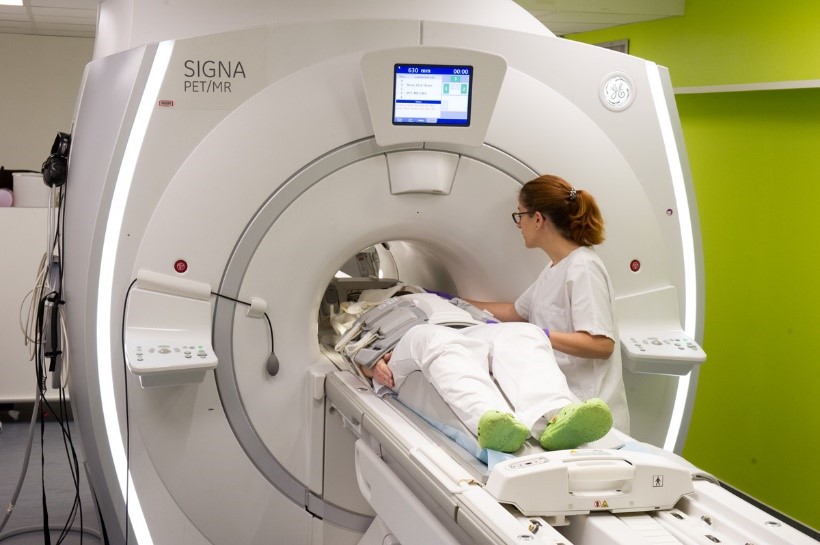

© L. Godart/CEA

These high-resolution imagers (2-3 mm) enable very precise visualization of brain tissue and vascular structures, thus contributing to accurate image quantification.

Moreover, the PET/MR (3T) camera, still rare in France, allows simultaneous acquisition of PET images and MRI sequences with enhanced sensitivity for improved image quality and reconstruction.

The high spatial resolution of the clinical cameras at the SHFJ allows a true continuum of PET projects initially carried out in MIRCen on preclinical cameras in animal models of neurodegenerative diseases (link to preclinical PET).

​​​

Example : PET images (co-registered with a MRI image) of a NHP (left) and a healthy subject (right) after injection of a tracer specifically targeting neuroinflammation